
Scientists Digitally Reconstruct the Face of ‘Little Foot,’ One of the Oldest Human Ancestors
Scientists have digitally reconstructed the face of ‘Little Foot,’ a 3.67-million-year-old Australopithecus fossil discovered in South Africa, offering new insights into early human evolution.
Mar 11, 2026

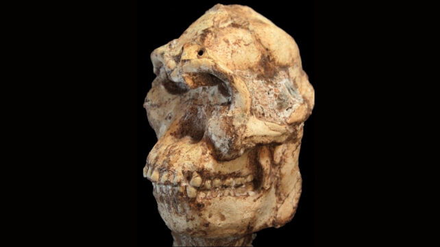
Scientists have digitally reconstructed the face of one of humanity’s oldest known ancestors, a fossil known as Little Foot, providing a clearer view of what early human relatives may have looked like nearly four million years ago. The fossil, formally catalogued as StW 573, belongs to an early member of the genus Australopithecus, a group of ancient hominins that lived in Africa millions of years before the emergence of modern humans.
Using advanced imaging technologies and computer modelling, researchers were able to reconstruct the crushed skull of the fossil and reveal its facial structure for the first time. The reconstruction represents a significant step in understanding early human evolution and the diversity of hominins that lived across Africa between four and three million years ago.
Discovery of the Little Foot Fossil
The fossil known as Little Foot was discovered in the mid-1990s in the Sterkfontein Caves near Johannesburg, South Africa, an area that forms part of the Cradle of Humankind, a UNESCO World Heritage Site famous for its numerous hominin fossils. The specimen was first identified in 1994 when paleoanthropologist Ronald J. Clarke discovered four small foot bones in a box of fossils at the University of the Witwatersrand. These bones indicated that the individual was capable of walking upright, leading to the nickname “Little Foot.”
Further investigation revealed that the rest of the skeleton was still embedded in rock within the Sterkfontein cave system. Excavating the fossil proved to be an extremely complex task because the bones were trapped in hard, concrete-like rock. Researchers spent nearly 15 to 20 years carefully extracting the skeleton from the cave deposits. The effort ultimately produced one of the most remarkable discoveries in paleoanthropology. Around 90 percent of the skeleton was recovered, making Little Foot the most complete Australopithecus skeleton ever found. This level of preservation allows scientists to study body proportions, movement patterns, and anatomical features in unprecedented detail.
The Timeline of Little Foot
Radiometric dating and geological analysis estimate that Little Foot lived around 3.67 million years ago, placing it among the oldest known hominin fossils discovered in southern Africa. Australopithecus species lived across Africa roughly between 4.2 million and 2 million years ago, and they represent an important stage in the evolutionary path that eventually led to the genus Homo, which includes modern humans. These hominins displayed a mixture of ape-like and human-like traits, such as the ability to walk upright while still retaining adaptations for climbing trees.
Little Foot’s skeletal structure suggests similar characteristics. Studies indicate that while it likely walked on two legs, it also had physical features that would have allowed it to climb trees, an ability that may have helped it escape predators such as large prehistoric cats.
Challenges in Studying the Skull
Although most of the skeleton was well preserved, Little Foot’s skull presented a major challenge for scientists. Over millions of years, geological pressure from cave sediments crushed and distorted the bones of the skull and face. This deformation made it impossible to reconstruct the skull using traditional physical methods without risking damage to the fossil.
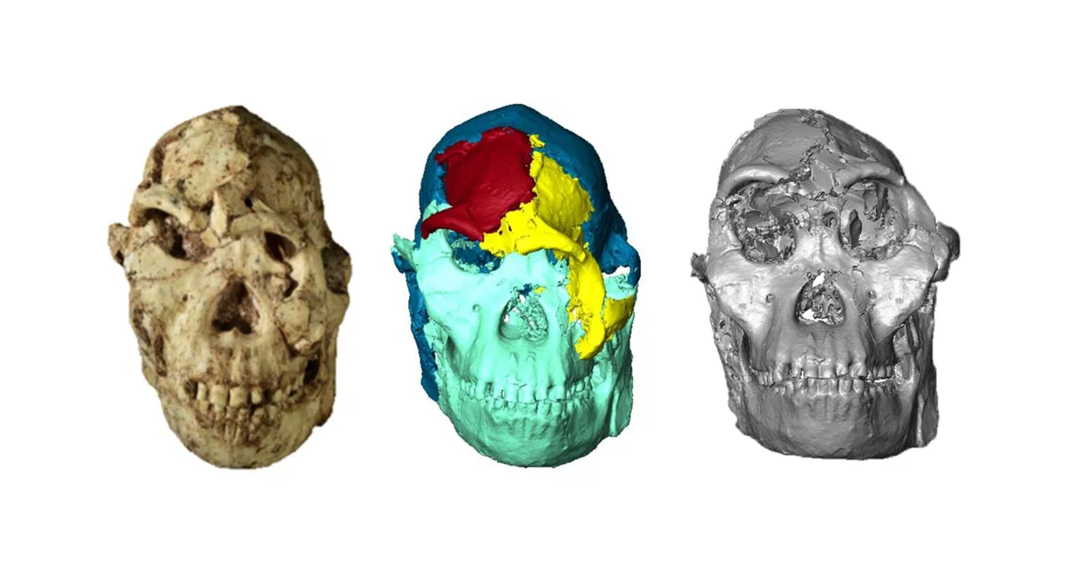
To overcome this problem, researchers turned to digital technologies. In 2019, the fossil skull was scanned using high-resolution X-ray micro-CT imaging at the Diamond Light Source synchrotron facility in the United Kingdom. The scans captured extremely detailed images of the internal and external structures of the bones, at resolutions as small as 21 micrometers.
Using these digital scans, scientists created a three-dimensional model of the skull and then used computer algorithms to virtually reposition the crushed bones back into their original anatomical positions. This process allowed researchers to reconstruct the facial structure without physically altering the fragile fossil.
The First Digital Face Reconstruction
The reconstruction work was led by paleoanthropologist Amélie Beaudet and an international team of researchers. Their study, published in the scientific journal Comptes Rendus Palevol, produced the first detailed reconstruction of Little Foot’s face.
The reconstructed face revealed several key features of this ancient hominin. The skull had relatively large eye sockets, and the facial proportions were found to fall between those of a gorilla and an orangutan in size, though the overall shape more closely resembled that of orangutans and bonobos.
Researchers also observed similarities between Little Foot’s facial anatomy and fossils discovered in East Africa. In particular, the structure around the eye sockets, known as the orbital region, shows resemblance to other Australopithecus specimens found thousands of kilometers away. These comparisons suggest that early hominin populations across Africa may have been more interconnected than previously thought. Some scientists propose that ancient populations may have migrated between eastern and southern Africa millions of years ago.
Scientific Significance
Little Foot is considered one of the most important fossils in the study of human evolution because of its completeness and age. Only a small number of Australopithecus fossils include well-preserved facial structures, making Little Foot an especially valuable specimen for anatomical comparison.
The reconstructed face also provides insights into key biological functions. According to researchers, the skull preserves anatomical regions involved in vision, breathing, and feeding, allowing scientists to study how these systems evolved in early human ancestors.
Furthermore, Little Foot may represent a unique or poorly understood species within the Australopithecus group. Some researchers suggest it may belong to Australopithecus prometheus, while others argue it could represent a previously unidentified lineage of early hominins. The debate highlights the complexity of the human evolutionary tree and the limited number of fossils available from this period.
Insights into Early Human Evolution
The digital reconstruction of Little Foot contributes to broader efforts to understand how early hominins evolved across Africa. By comparing its anatomy with other fossils, scientists can explore questions about migration, adaptation, and the diversification of early human ancestors.
Because Little Foot lived during a time when different hominin species were evolving across the continent, its anatomy provides a valuable reference for studying variation within the Australopithecus genus. The reconstruction also demonstrates how modern imaging technology can reveal new information from fossils that have been known to science for decades.
Researchers plan to continue analyzing other parts of the skull, including the teeth and braincase, using similar digital methods. These studies may reveal additional details about diet, sensory capabilities, and brain evolution in early human relatives.